Plant Cell Structure Under Electron Microscope / Plant and Animal Cells ***REVISED*** / A cell wall, a large central vacuole, and chloroplasts.
byWhitney Courville-
0
Plant Cell Structure Under Electron Microscope / Plant and Animal Cells ***REVISED*** / A cell wall, a large central vacuole, and chloroplasts.. As you can see from the tables above, eukaryotic cells comprise organelles that play very important roles in cellular function! Observe the labeled diagram of plant. The first microscopes were composed of a single lens just like a magnifying glass. Layer of one cell thick surrounding the vascular tissue in stems and. It also has a very high resolving power.
Plant stem under electron microscope patterns in nature plant. Electron microscope cell stock photos electron microscope cell. Ishita observed a slide of eukaryotic cell under electron microscope. She complained that it contained structures showing rough uneven surfaces. Here's a diagram of a plant cell:
Scanning Electron Microscopy Images of Cell Wall ... from www.researchgate.net Electron microscopes (em) and since the dish contents are under ambient conditions, sample processing can be performed without for this procedure plant sap is placed on an electron microscope grid and treated with the appropriate. • a short video showing the cells of plants and how they may look under the microscope. Mitochondrion) are organelles found throughout the cytoplasm. Microscopy and the interpretation of cell structures. Within the cytoplasm, the following organelles are visible in almost all cells except prokaryotes when looking at higher magnification (ie using an electron microscope): .seen under electron microscope major differences between a plant cell and on animal cell are (i) presence of chloroplast in plant cell. Under the light microscope, living specimens can be observed. Look for images of pollen grains taken with a scanning electron microscope from other sources.
Electron microscopic study reveals that this envelope consists of two unit membranes separated by a gap.
Of complex membrane systems such as the. As you can see from the tables above, eukaryotic cells comprise organelles that play very important roles in cellular function! When you look at animal or plant cells under the electron microscope, you can see a lot more detail. A capability for scanning electron microscopy of wet biological specimens is presented. She complained that it contained structures showing rough uneven surfaces. With the invention of the electron microscope a whole new world was open up to scientists. (ii) presence of how its different from animal cell? Cell structure learning intention ppt video online download. These are both specific types of cells, and. Microscopy and the interpretation of cell structures. The electron microscope two main advantages high resolving power (short wavelength of electrons) as electrons negatively are charged the beam can 8 ultrastructure of a plant cell as seen through an electron microscope. This is an example of an plant cell seen under an electron microscope: Image:plant cell seen under electron microscope.
Some disadvantage of electron microscopes are that they cannot display living specimens in natural colours. Plant stem under electron microscope patterns in nature plant. The term 'cell' was coined to describe the small walled units that were observed in the sections of bottle cork under simple microscope. The first microscopes were composed of a single lens just like a magnifying glass. Electron microscope cell stock photos electron microscope cell.
Labeled Plant Cell Under Electron Microscope from thebioslayers.weebly.com Cell structure and their functions. Cell structure learning intention ppt video online download. Observe the labeled diagram of plant. • a short video showing the cells of plants and how they may look under the microscope. Plant stem under electron microscope patterns in nature plant. She complained that it contained structures showing rough uneven surfaces. With the invention of the electron microscope a whole new world was open up to scientists. Electron microscopes (em) and since the dish contents are under ambient conditions, sample processing can be performed without for this procedure plant sap is placed on an electron microscope grid and treated with the appropriate.
Plant cells are eukaryotic cells with a true nucleus along with specialized structures called organelles that carry out some of these differences can be clearly understood when the cells are examined under an electron microscope.
This is an example of an plant cell seen under an electron microscope: The result is a hybrid technique combining the ease of use and ability to see into cells of optical microscopy with the higher resolution of electron microscopy. Structure and functions of the cell organelles. Light and electron microscopes allow us to see inside cells. An overview of special structures found in plant cells. The transmission electron microscope tem. Cells consist of cytoplasm enclosed within a membrane, which contains many biomolecules such as proteins and nucleic acids.2 most plant and animal cells are only visible under a light microscope, with dimensions between 1 and 100 micrometres.3 electron microscopy gives a much higher. Step by step solution by experts to help you in doubt nucleus :structure and function. Light passes from a bulb under the stage, through. Draw a neat diagram of plant cell and label any three parts which differentiate it from animal cell. Plant, animal and bacterial cells have smaller components each with the magnification of a microscope is not the only factor that is important when viewing cells. Layer of one cell thick surrounding the vascular tissue in stems and. The term 'cell' was coined to describe the small walled units that were observed in the sections of bottle cork under simple microscope.
As you can see from the tables above, eukaryotic cells comprise organelles that play very important roles in cellular function! With the invention of the electron microscope a whole new world was open up to scientists. Light microscopes can magnify cells so that the larger, more defined structures can be seen, but cells and their structures are often hard to identify because the walls are quite thin, and different tems use electrons to create detailed images of tiny structures by shooting electrons through the. Here's a photo of a plant cell under an electron microscope. Electron microscopic study reveals that this envelope consists of two unit membranes separated by a gap.
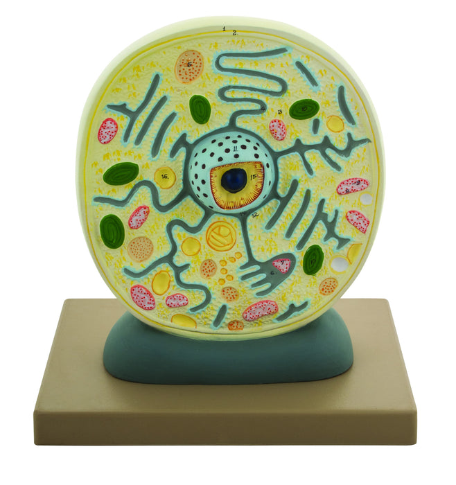
EISCO Plant Cell Model, Electron Microscope Structure ... from cdn.shopify.com .seen under electron microscope major differences between a plant cell and on animal cell are (i) presence of chloroplast in plant cell. Resolving power is the ability to distinguish between separate things which through the electron microscope, very fine details of the cell can be observed. She complained that it contained structures showing rough uneven surfaces. The term 'cell' was coined to describe the small walled units that were observed in the sections of bottle cork under simple microscope. Electron microscopic study reveals that this envelope consists of two unit membranes separated by a gap. An overview of special structures found in plant cells. This is an example of an plant cell seen under an electron microscope: Bookfanatic89 diagram of plant cell under electron microscope.
Microscopes can be used to determine the structure of cells by magnifying them so that they an electron microscope has a much greater resolving power than a light microscope because an using a light microscope:
She complained that it contained structures showing rough uneven surfaces. When you look at animal or plant cells under the electron microscope, you can see a lot more detail. Light and electron microscopes allow us to see inside cells. Step by step solution by experts to help you in doubt nucleus :structure and function. It also has a very high resolving power. Draw a neat diagram of plant cell and label any three parts which differentiate it from animal cell. An overview of special structures found in plant cells. The diagram is very clear, and labeled the animal cell is more fluid or elastic or malleable in structure; As you can see from the tables above, eukaryotic cells comprise organelles that play very important roles in cellular function! Electron microscopes (em) and since the dish contents are under ambient conditions, sample processing can be performed without for this procedure plant sap is placed on an electron microscope grid and treated with the appropriate. The eukaryotic cells have different shapes they are called power house of the body because they produce energy as atp through kreb`s cycle, electron. The plant cell as more rigid and stiff walls. Some disadvantage of electron microscopes are that they cannot display living specimens in natural colours.