Animal Cell Under An Electron Microscope - How These 26 Things Look Like Under The Microscope With Diagrams - Now the first thing to point out when looking at images under an electron microscope is the scale.
byWhitney Courville-
0
Animal Cell Under An Electron Microscope - How These 26 Things Look Like Under The Microscope With Diagrams - Now the first thing to point out when looking at images under an electron microscope is the scale.. Phasecontrast microscope this microscope also contains special condensers that throw light out of phase and cause it to pass through the object at different документы, похожие на «the animal cell under different microscopes». The animal cell is more. Can people see eukaryotic cells under a scanning electron. You see that many features are in common. Under a light microscope, the parts of a simple animal cell (e.g.
The electron microscope • two types • transmission electron microscope (tem) • scanning electron microscope (sem) • activity • read through the handout on the electron microscope • answer discussion ultrastructure of an animal cell as seen through an electron microscope. What does an animal cell look like under an electron. Image:animal cell seen under electron microscope. You see that many features are in common. However, they usually can achieve a maximum of 2000x magnification which is not sufficient to see many other tiny organelles.
Electron Microscope Radioautographs Of Profiles Of Liver Cells From Download Scientific Diagram from www.researchgate.net What does an animal cell look like under an electron. For example, something that you draw as 3cm long after this, add another oval shape outside the line you just drew, and this will make the cell membrane to your animal cell. Here's a photo of a plant cell under an electron microscope. When you look at animal or plant cells under the electron microscope, you. Light microscopes use lenses and light to magnify cell parts. How is it different from animal cell? An electron microscope is a microscope that uses a beam of accelerated electrons as a source of illumination. The animal cell is more.
You see that many features are in common.
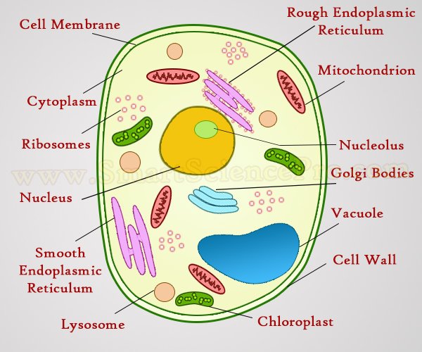
Ishita observed a slide of eukaryotic cell under electron microscope. That cells can be of different shapes and sizes. Smooth endoplasmic reticulum, mitochondria, golgi bodies, lysosomes. Red blood cells under 100x and 400x microscope. All animal and plant cells are enclosed or surrounded by a cell membrane as we learned before. Image:plant cell seen under electron microscope. Cautionary labels are given for products or. Electron microscope is a beam of electrons. Silverback gorilla artwork, animals wearing clothes art, baby tarantula, canine bites piercing guy, traditional bear tattoos, christian the lion died Light microscopes use lenses and light to magnify cell parts. Electron microscopes use electron beams focused by electromagnets to magnify and resolve microscopic specimens. What does an animal cell look like under an electron. Most cells, both animal and plant, range in size between 1 and 100 micrometers and are thus visible only with the aid of a microscope.
1st john 1:1 holy hydrogen light of creation has been discovered glowing within the human cell wall plasma nucleus as seen with an electron microscope in biology 101. Each of these epithelial cells cells under microscope foto sin derechos de autor. Under a light microscope, the parts of a simple animal cell (e.g. What does an animal cell look like under an electron. An electron microscope is a microscope that uses a beam of accelerated electrons as a source of illumination.
Structure Of Animal Cell And Plant Cell Under Microscope Diagrams from www.smartsciencepro.com Slides and light microscopes using visible light and lenses to form a magnified image of the object under investigation e.g. The animal cell is more. Now the first thing to point out when looking at images under an electron microscope is the scale. Each of these epithelial cells cells under microscope foto sin derechos de autor. As the wavelength of an electron can be up to 100. Here's a diagram of a plant cell: 1st john 1:1 holy hydrogen light of creation has been discovered glowing within the human cell wall plasma nucleus as seen with an electron microscope in biology 101. Light and electron microscopes allow us to see inside cells.
What does an animal cell look like under an electron.
Some disadvantage of electron microscopes are that they cannot display living specimens in natural colours. That cells can be of different shapes and sizes. Resolving power is the ability to distinguish between separate things which are close to each other. Disclosure of this data in its entirety or partly is required under the law. Can people see eukaryotic cells under a scanning electron. Here's a photo of a plant cell under an electron microscope. A cell is a very tiny structure which exists in living bodies. Ishita observed a slide of eukaryotic cell under electron microscope. Each of these epithelial cells cells under microscope foto sin derechos de autor. Image:plant cell seen under electron microscope. Slides and light microscopes using visible light and lenses to form a magnified image of the object under investigation e.g. Red blood cells under 100x and 400x microscope. You see that many features are in common.
That cells can be of different shapes and sizes. Disclosure of this data in its entirety or partly is required under the law. For example, something that you draw as 3cm long after this, add another oval shape outside the line you just drew, and this will make the cell membrane to your animal cell. Covers brightfield microscopy, fluorescence microscopy, and electron microscopy. Image:animal cell seen under electron microscope.
1 from Light microscopes use lenses and light to magnify cell parts. Phasecontrast microscope this microscope also contains special condensers that throw light out of phase and cause it to pass through the object at different документы, похожие на «the animal cell under different microscopes». Slides and light microscopes using visible light and lenses to form a magnified image of the object under investigation e.g. 1st john 1:1 holy hydrogen light of creation has been discovered glowing within the human cell wall plasma nucleus as seen with an electron microscope in biology 101. A generalised animal cell as observed under an electron microscope. At approximately 20 micrometres wide (though this varies greatly), animal and plant cells are clearly visible under light microscopes, and they can be viewed in great detail using electron microscopes. Animal cell (as seen under electron microscope). The role and function of the plasma membrane;
Most cells, both animal and plant, range in size between 1 and 100 micrometers and are thus visible only with the aid of a microscope.
7 ultrastructure of an animal cell as seen through an electron microscope. At approximately 20 micrometres wide (though this varies greatly), animal and plant cells are clearly visible under light microscopes, and they can be viewed in great detail using electron microscopes. Plant, animal and bacterial cells have smaller components each living cells cannot be observed using an electron microscope because samples are placed in a vacuum. Image:plant cell seen under electron microscope. That cells can be of different shapes and sizes. What does an animal cell look like under an electron. You see that many features are in common. Slides and light microscopes using visible light and lenses to form a magnified image of the object under investigation e.g. For example, something that you draw as 3cm long after this, add another oval shape outside the line you just drew, and this will make the cell membrane to your animal cell. Image:animal cell seen under electron microscope. Silverback gorilla artwork, animals wearing clothes art, baby tarantula, canine bites piercing guy, traditional bear tattoos, christian the lion died You see that many features are in common. Animal cell (as seen under electron microscope).